The doctor enters the operating room, dons a Virtual Reality helmet and, while observing an exact reproduction of the organ, in three dimensions, initiates the operation, using a computer that guides him at all times. This sounds like something out of a sci-fi movie, but has already been done at more than one hospital. What once seemed a fantasy a decade ago is now a fact, as Virtual Reality is already revolutionizing the world of surgery.
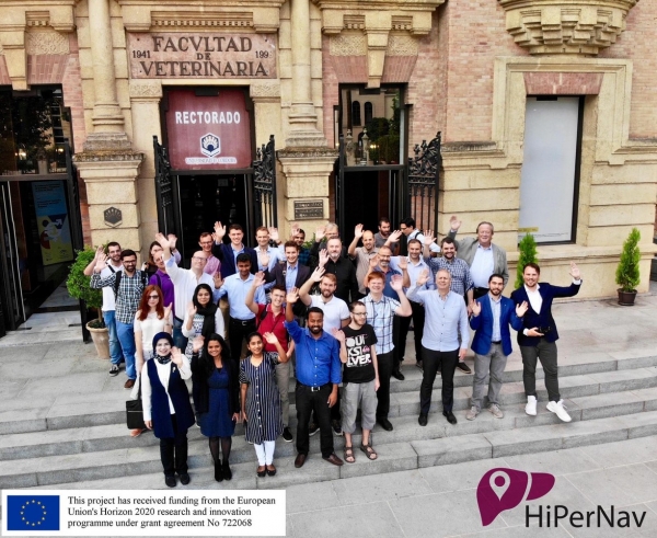
The HIPERNAV international research project got started three years ago with the aim of advancing automatic technology for the removal of soft tissue tumours. Specifically, research has focused on the ablation of liver cancer, one of the most frequent tumours with the highest mortality worldwide.
The initiative, comprised of experts in different branches of Medicine and Computing, has developed a new system, equipped with high-precision technology that is able to reduce the time required for this operation from four hours to just one. According to researcher Joaquín Olivares, head of the Advanced Computing Research Group at the University of Cordoba, and the only representative in Spain involved in the project, the system provides the doctor with a Virtual Reality helmet and a screen featuring an exact reconstruction of the patient's organ, in 3D. Once the organ is cut, the surgeon uses a screen with a target that turns green when the instruments are properly positioned on the tumour. Then, after activating a button, a machine performs the ablation, automatically, with total precision.
The system, which draws on the technological improvements that have been incorporated into the world of surgery in recent years, and the extensive experience of the members making up the project, began by testing on animals, and has already been validated in humans. In Norway, a hospital ward is being built for this type of intervention, and in Switzerland several operations have already been carried out using this technology with satisfactory results. The post-operative period is reduced to one or two days because it is a less invasive procedure and, as Olivares points out, "with this mechanism, less of the liver is lost, which also improves the patient's quality of life".
The University of Cordoba has worked on the processing of medical images, one of the fundamental parts of the project, which allows for the depiction of the organ and its vessels in three dimensions. Specifically, the Advanced Computing Research Group has provided high-performance computing techniques so that the processing can be done in real time and in adequate resolution.
The project, which is already beginning to work on pancreatic cancer, boasts great potential, with a view to the future. This type of technology requires a great amount of data, so the challenge, Olivares stressed, is to improve computing capacity in order to increase the resolution of the images. In this way it will be possible to augment the precision of the operations of tomorrow; a future in 3D that is bound to arrive, and in which doctors will supervise surgeries carried out by machines.


